上期回顧:
本綜述回顧了CT血管造影術(shù)的演變��,從其發(fā)展和早期挑戰(zhàn)到成熟產(chǎn)品,提供對(duì)于心血管疾病發(fā)現(xiàn)和治療獨(dú)有的視野����。
本期內(nèi)容:
CTA的起源,初期的瓶頸以及解決的方法
CTA的起源
Emergence of Angiography
CT血管造影(CTA)得以實(shí)現(xiàn)的主要技術(shù)支持是在1990年臨床上引入了螺旋(6) (7)掃描����,標(biāo)志著進(jìn)入容積CT時(shí)代。它的主要貢獻(xiàn)是取代了“步進(jìn)掃描”(step and shoot)的采集模式��。該模式是在一次橫斷位圖像采集的時(shí)候���,掃描床是靜止的��,而在下一次機(jī)架旋轉(zhuǎn)重啟掃描前��,掃描床前移至下一個(gè)掃描位置���。螺旋掃描是在掃描床進(jìn)動(dòng)中連續(xù)采集,可在單位時(shí)間內(nèi)覆蓋更大的掃描范圍��。
The primary enabling technology for CT angiography was the clinical introduction of spiral (6) or helical (7) scanning in1990, which ushered in the era of volumetric CT. Its key contribution was the replacement of the “step and shoot” acquisition mode, in which the table wasstatic during the acquisition of a single transverse section and subsequently advanced to the next scan position before gantry rotation resumed, with continuous acquisition of projections during table travel that allowed coverage of muchlarger volumes per unit time.
其他方面的優(yōu)勢(shì)是可以采集靜脈團(tuán)注對(duì)比劑的首過(guò)(動(dòng)脈期)��,當(dāng)它流經(jīng)特定的血管區(qū)域���。在早期���,床速受限于機(jī)架每轉(zhuǎn)的層厚,例如��,機(jī)架每轉(zhuǎn)1秒�����,層厚3mm����,則床速為3mm/秒。另一個(gè)限制就是X光球管熱容量��,它限定毫安量(也因此噪聲增加)���,及掃描時(shí)間小于30秒�,結(jié)果同樣的掃描參數(shù)下最大覆蓋范圍是9cm(8)����。
Among other advantages, this permitted the capture of the first pass of anintravenous contrast agent bolus as it transited a particular vascular territory. In those early days, table speed was limited to the section thickness per gantry rotation; thus, for example, given a 1-second gantry rotation and 3-mm sections, table speed was 3 mm/sec. Another limitation wasx-ray tube heat capacity, limiting milliamperage (and, thereby, in- creasingnoise) and scan time to less than 30 seconds, resulting in a maxi- mum coverage with the same parameters of 9 cm (8).
第一篇描述CT血管造影(CTA)的文章發(fā)表在1992年11月的放射學(xué)(Radiology)雜志上(9,10),并且展示了快速容積覆蓋與3D可視化的可能性。
The first articles describing CT angiography appeared in the November 1992edition of Radiology (9,10) and demonstrated the possibilities givenfast volume coverage and 3D visualization.
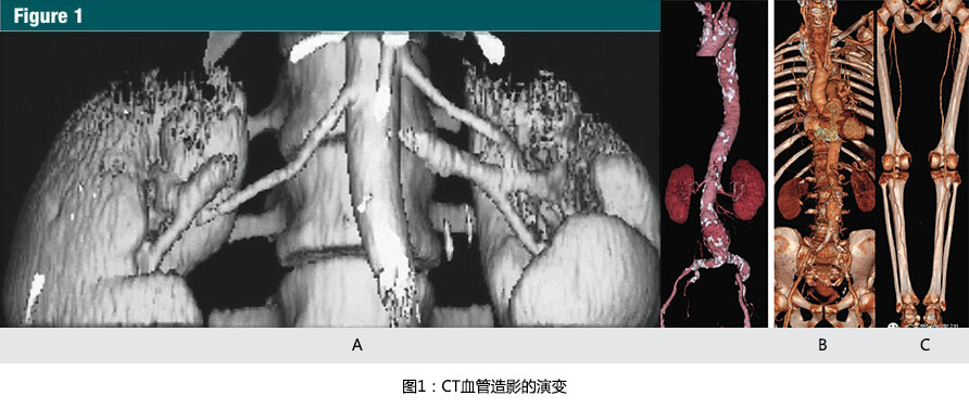
在1991年到1998年��,由于單排螺旋CT的掃描速度限制了CTA進(jìn)入到各個(gè)不同的血管領(lǐng)域�����。采用大于1的掃描螺距之前�,一個(gè)層厚3mm,30秒的掃描��,最大覆蓋的范圍是9cm����,因此,限制了在頸外動(dòng)脈(10)���、Willis環(huán)(11)���,腎動(dòng)脈(12,13)(圖 1a)及近端腹主動(dòng)脈(13)中的早期應(yīng)用�。
From 1991 to 1998, single-detector row spiral CT technique limitedclinical CT angiography to discrete vascular territories. Prior to the introduction of scan pitch values greater than one, a scan with 3-mm nominalsection thickness provided a maximum of 9 cm table travel in 30 seconds andthus limited initial applications to the extracranial carotid arteries (10),the circle of Willis (11), the renal arteries (12,13)(Fig 1a), and the proximal abdominal aorta(13).
而3mm層厚是最多用于多平面重建(MPR)與3D容積再現(xiàn)(VR)重建,早期的CTA研究者能有效地利用螺旋CT5mm層厚重建的橫斷面圖像來(lái)評(píng)價(jià)中心肺動(dòng)脈(14)與胸主動(dòng)脈(15)��。
While a nominal section thickness of 3 mm was considered maximal for thecreation of useful multiplanar reformations and 3D renderings, early CT angiography pioneers effectively used the primary transverse reconstructions from spiral CT acquisitions with 5 mm thickness to evaluate the central pulmonary arteries (14)and the thoracic aorta (15).
隨著臨床引入1~2之間的掃描螺距(16)�,最大的解剖覆蓋范圍增加了1倍,為擴(kuò)大其臨床應(yīng)用鋪平了道路,包括急性主動(dòng)脈綜合征(AAS)的評(píng)價(jià)��、創(chuàng)傷患者主動(dòng)脈損傷的探查及制定動(dòng)脈瘤治療計(jì)劃的關(guān)鍵環(huán)節(jié)-主動(dòng)脈定性和定量分析�����。
With the clinical introduction of scan pitch values between one and two (16), maximal anatomic coverage doubled, paving the way for an expansion in clinicalapplications, to include assessments of acute aortic syndromes (AAS), the detection of aortic injury in trauma patients, and the qualitative and quantitative characterization of the aorta as key enablers for planning aneurysm therapy.
隨著研究關(guān)注在CT血管造影(CTA)掃描技術(shù)和后處理的提升����,新的CTA的臨床應(yīng)用可以拓展到整個(gè)血管內(nèi)腔�、管壁以及終末器官的容積重建(14,17-22)�。
While many investigations focused on the refinement of the techniques for CT angiogram acquisition and post-processing, new clinical insights were made possible by CT angiography’s ability to volumetrically resolve the entirety ofthe blood vessel lumen, wall, and end organ (14,17–22).
隨著CTA臨床應(yīng)用的潛力,行業(yè)尋求進(jìn)一步(更大)容積覆蓋通過(guò)更快機(jī)架旋轉(zhuǎn)及每轉(zhuǎn)可采集更多層面的多排并行探測(cè)器的開(kāi)發(fā)����。
With the clinical potential for CT angiography established, the industry sought to further increase volume coverage through the development of scanners with faster gantry rotation and with multiple parallel detector rings that acquired more than one section per rotation.
1998年推出的早期多排探測(cè)器CT具有4排探測(cè)器和0.5s的旋轉(zhuǎn)時(shí)間,對(duì)于相同層厚����,單位時(shí)間增加了8倍的容積覆蓋范圍(8,23)(圖 1b�,1c)。
Early multi-detector row CT scanners introduced in 1998 had four detector rings and were capable of 1/2-second gantry rotations, effectively multiplying volume coverage per unit time 38 at the same section thickness (8,23)(Fig 1b, 1c).
15年后����,今天多排探測(cè)器CT已經(jīng)發(fā)展到320排探測(cè)器����,機(jī)架旋轉(zhuǎn)時(shí)間最快達(dá)到270ms�,并且在一些案例中雙源CT允許在最多數(shù)秒內(nèi),進(jìn)行亞毫米各項(xiàng)同性的大范圍容積采集���。
Fifteen years later, today's multi-detector CT scanners have up to 320 detector rings, gantry rotation times as low as 270 msec, and in some cases twox-ray sources, allowing sub-millimeter isotropic resolution to be acquired oververy large volumes in, at most, a few seconds.
快速容積覆蓋還能極大地減少對(duì)比劑的使用量���,而無(wú)明顯的血管丟失(24)。
Faster volume coverage also allowed a sizable reduction in contrast media usage without loss in vascular conspicuity (24).
隨著多排探測(cè)器CT進(jìn)入(臨床)�,幾乎所有長(zhǎng)軸方向覆蓋范圍的限制都消失了,為CT血管造影(CTA)在下肢動(dòng)脈系統(tǒng)(25)�,全頭頸部血管系統(tǒng)(23),胸�����、腹主動(dòng)脈系統(tǒng)(23)����,和上肢動(dòng)脈(26)系統(tǒng)的成像鋪平了道路。
With the introduction of multi-detector CT, virtually all limits on longitudinal coverage disappeared, paving the way for CT angiography to beapplied to imaging the inflow and run-off of the lower extremity arterialsystem (25), the entire cervicocranial vascular system (23), thethoracoabdominal aortoiliac system (23), and the upper extremity arterial system (26).
到2002年����,最后一個(gè)動(dòng)脈系統(tǒng)-冠狀動(dòng)脈成像依然存在問(wèn)題��。然而采用4排與8排CT的冠脈血管造影的概念驗(yàn)證調(diào)查發(fā)表(27)��,16排冠脈CT血管造影(CTA)進(jìn)入臨床實(shí)踐�,伴隨64排CT在2005年誕生冠脈血管造影(CTA)成為了主流���。
By the year 2002, there was but one final arterial frontier remaining—the coronary arteries. While proof-of-concept investigations of coronary CT angiography were published using four- and eight-row multi-detector CT scanners(27), the introduction of 16-row multidetector CT brought coronary CT angiography to clinical practice and with 64-row multi-detector CT in 2005 it became mainstream.
計(jì)算機(jī)與圖像后處理
Computersand Image Processing
高速的CT的飛速演變與摩爾定律保持一致,它預(yù)示著晶體管集成化的密度大約每2年倍增����。
The rapid evolution of fast CT scanners is congruent with Moore's law,which predicts the doubling of the density of transistors on integrated circuits approximately every 2 years.
除了影響采集電路系統(tǒng),摩爾定律還適用于計(jì)算機(jī)性?xún)r(jià)比的快速增加�,而沒(méi)有用于重建高分辨錐形束容積采集的費(fèi)用和時(shí)間的增加,這由許許多多層面組成��,這些是沒(méi)有臨床實(shí)用性的���。
In addition to effecting the acquisition circuitry, Moore’s law is also responsible for the rapid increase in computer performance/price ratios,without which the expense and time to reconstruct these high-resolutioncone-beam volume acquisitions, consisting of hundreds to thousands of sections, would not be clinically practical.
摩爾定律也直接成為CT血管造影(CTA)最終臨床應(yīng)用的推動(dòng)者:因?yàn)橐粚咏又粚拥腃TA圖像并不有效和直觀���,可視化的CT血管造影包括表面遮蓋顯示,最大密度投影����,和容積再現(xiàn)(VR)(圖1b�,1c)����。
Moore's law is also directly responsible for the final enabler of clinicalCT angiography: Because section-by-section inspection of CT angiographic imagesis neither efficient nor intuitive, visualization of CT angiography studies employs shaded surface displays, maximum intensity projections, and volume rendering (Fig 1b, 1c).

(A):1991年12月獲得的腎動(dòng)脈CT血管造影圖像。9厘米的縱向覆蓋���,采用3mm的準(zhǔn)直線束需要30秒螺旋掃描時(shí)間��。當(dāng)時(shí)����,表面遮蓋技術(shù)是唯一的三維顯示手段�����。最大密度投影和容積再現(xiàn)成像需要在高度專(zhuān)業(yè)的計(jì)算機(jī)系統(tǒng)上進(jìn)行脫機(jī)處理(參考文獻(xiàn)13)
(B):隨著1998年的四排螺旋CT的引進(jìn)�����,使主動(dòng)脈-髂動(dòng)脈系統(tǒng)(從胸廓入口開(kāi)始直到腹股溝)作為一個(gè)整體僅通過(guò)一次圖像采集并成像成為了可能���。容積再現(xiàn)技術(shù)所展示的CTA圖像����,使用4x2.5mm的螺旋掃描模式在28秒內(nèi)掃描完成。圖片充分顯示了主-髂動(dòng)脈鈣化和腹主動(dòng)脈瘤(參考文獻(xiàn)8)
(C):2001年使用容積再現(xiàn)技術(shù)展示的CTA圖像�,使用16 x1.25毫米螺旋掃描模式,僅用了21秒的時(shí)間����,就完成了從顱底至踝的動(dòng)脈系統(tǒng)掃描,離1991年第一例螺旋CT的CTA成像只隔了10年��,但是CT的掃描速度則增加了近25倍�����。
然而����,早期數(shù)據(jù)集僅包含數(shù)十層截面����,每個(gè)所需的視圖方向需要很長(zhǎng)時(shí)間在昂貴的工作站上來(lái)計(jì)算完成。
While early datasets consisted of only tens of cross-sections, each of themany desired view directions required many seconds to compute on expensive workstations.
今天����,許多供應(yīng)商都可以提供在廉價(jià)計(jì)算機(jī)上運(yùn)行的軟件�,基于數(shù)千幅增強(qiáng)CT斷層圖像的高級(jí)光影效果���,這些軟件可以獲得交互式的高分辨率容積再現(xiàn)(VR)���,如自動(dòng)去骨和曲面重建。
Today, many vendors provide software that runs on inexpensive computersand is capable of inter- active high-resolution volume rendering with advanced lighting effects based on thousands of contrast-enhanced CT sections withadditional capabilities such as automated bone removal and curved planar reformatting.
CTA數(shù)據(jù)分析已經(jīng)發(fā)展成這種模式:先經(jīng)專(zhuān)用的后處理解決方案進(jìn)行定制的可視化和定量分析�����,然后再行橫斷面重建進(jìn)行分析���。
The analysis of CT angiographic datasets has evolved to the point where review of the transverse reconstructions is a secondary analysis to tailored visualization and quantitation tasks using application-specific post-processing solutions.


